 log
log My
My Contact


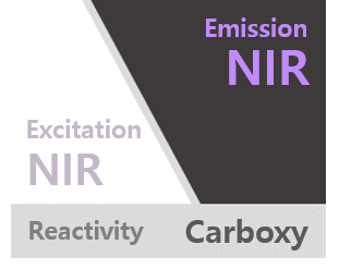
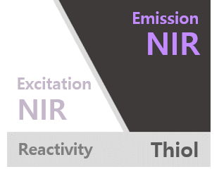
ICG Thiol
Catalog No. PWT1301
| PACKING UNIT | price |
Lead time |
|---|---|---|
| 1 mg | $255.00 | |
| 5 mg | $913.00 | |
| 25 mg | $3,396.00 |
Description
ICG Thiol is an inactive form of near infrared (NIR) fluorescent dye and used to generate a stable fluorescence signal in bioimaging. The thiol is attached to ICG fluorophore trough a spacer. NIR fluorescence allows to observe the deep image from the surface of skin and being utilized in a wide range of research fields. The maxima of Ex/Em values are at 785/812 nm. ICG might be excited using 750-800 nm laser line or LED and displays excellent optical property. ICG thiol can be labeled to biomolecules through a disulfide bond formation with thiol of cysteine residue or can be utilized as a reference standard for dye-conjugates.
Citation & Reference
1. Masashi Gotoh. Development of a canine model of pulmonary emphysema and imaging of the emphysematous lung with infrared thoracoscopy. J Thorac Cardiovasc Surg 126.6 (2003): 1916-21.
2. Aaron M. Mohs. An integrated widefield imaging and spectroscopy system for contrast-enhanced, image-guided resection of tumors. IEEE Trans Biomed Eng 62.5 (2015): 1416-24.
3. Mohammed Hassan. Near Infrared Fluorescence Imaging with ICG in TECAB Surgery Using the da Vinci Si Surgical System in a Canine Model. J Card Surg 27.2 (2012): 158-162.
4. R. C. Benson. Fluorescence properties of indocyanine green as related to angiography. Phys Med Biol 23.1 (1978): 159-63.
5. Mitsuharu Miwa. The Principle of ICG Fluorescence Method. The Open Surgical Oncology Journal 2 (2010): 26-28.
OPTION
Total
$0


Added cart
We put the items
in the shopping cart.
