BioActs offers the full spectrum of fluorescent dyes such as FSD FluorTM, Flamma® Fluors, ICG, and Other Dyes,
and also offers Fluorescent Quenchers, Crosslinkers, Fluorescent Antibodies, Bioprobes,
and Microspheres, Magnetic Beads, Dye Labeling Kit, etc.
In Vivo Imaging Probes (Fluorescence)
NpFlamma® HGC series
NpFlamma®
HGC series is a fluorescent dye incorporated biocompatible and biodegradable
chitosan based amphiphilic nanoparticle that enables to selectively detect
tumor cells. Chitosan nanoparticles can selectively accumulate in cancer tissues due to high permeability for
loose new blood vessels around cancer tissues and retention effect. The
polymeric nanoparticles form self-aggregates size of several hundred nanometers
in the aqueous system accumulate only in vicinity of cancer tissues. Chitosan
particles display low toxicity along with absence of
noticeable side effect in vivo, yet
they exhibit a long half-life, high stability and aqueous solubility. Thus,
NpFlamma® HGC series is an ideal fluorescence agent for in vivo imaging of angiography and tumor
progression. In conjunction with NIR dyes, the series allows to observe
non-invasive images of cancer metastases and to contrast blood vessel. The
hydrophobic nature of NpFlamma® HGC series can embed hydrophobic
materials, thus they can be utilized as a selective carrier for hydrophobic
cancer drugs such as doxorubicin and paclitaxel, etc. In addition, carboxylic
acids on the surface of NpFlamma® HGC series can bind to a variety of
biomolecules via amide or ester linkages enabling to utilize them in
multi-purposes.
NpFlamma® MMP series
Matrix metalloproteinases (MMPs) are a group of calcium-dependent, zinc-containing endopeptidases that that in concert are responsible for the degradation of most extracellular matrix (ECM) proteins during organogenesis, growth and normal tissue turnover. Humans have 23 MMPs family, and the expression and activity of MMPs in adult tissues is normally quite low, but increases significantly in various pathological conditions that may lead into unwanted tissue destruction, tumor growth and metastasis, inflammatory diseases, etc. Cancer cells secrete VEGF and FGF in order to induce angiogenesis in vascular endothelial cells, but cannot reach to vascular endothelial cells as being caught in ECM. Although MMPs are connected with cancer cells survival and expansion, only very small amount of MMPs are synthetized by cancer cell. However, the cancer cells induce inflammatory cells to secrete MMPs for assisting the delivery of VEGF secreted by cancer cells and make peripheral space for angiogenesis. The connection between MMPs and apoptosis, cell migration and angiogenesis enables to utilizing MMPs as tumor markers. An increased expression and activity of MMPs in both tissues and blood of patients with various types of cancer is observed.
BioActs developed NpFlamma® MMP series, MMP-activatable polymeric
in vivo fluorescent probes, for early diagnosis and for visualization of
overexpressed MMPs related diseases. The probes consist of a fluorescent dye
that connected to a quencher through a MMP-cleavable peptide, and a chitosan
based nanoparticle (CNP). The one end of peptide is chemically conjugated to a
chitosan based nanoparticle. NpFlamma® MMPs
are optically silent in
their inactivated state and become highly fluorescent following MMP-cleaved
activation. CNP can
selectively accumulate in cancer tissues due to high permeability for loose new
blood vessels around cancer tissues and retention effect. The polymeric
nanoparticles form self-aggregates size of several hundred nanometers in the
aqueous system accumulate only in vicinity of cancer tissues. Thus, CNP enables to bring up the probe to tumor cells, and the cleavage
of the peptide by MMPs allows to selective detection of tumor by realizing
fluorescence imaging.
NpFlamma®
MMP series equipped with several different MMPs (MMP-2, -3, -7, -9, -13)
cleavable peptides that enable to detect a wide range of diseases. Since self-assembled CNPs
have already been used as vehicles for hydrophobic drug delivery, and have
shown therapeutic efficacy for mouse tumors. Therefore, the role of NpFlamma®
MMP series might be extended as theranostic agents that simultaneously
monitoring therapeutic responses and delivering therapy. BioActs offers
NpFlamma® MMP series as smart fluorescent probes for monitoring
MMP-related diseases such as cancer progression, invasion and metastasis,
rheumatoid arthritis, pulmonary diseases and areas of cardiovascular disease,
and also for evaluating the potential therapeutic efficacy of drugs targeting
for these diseases.
AngioFlamma® series
Integrins are heterodimeric transmembrane receptors for cell adhesion to extracellular matrix (ECM) proteins and play important roles in certain cell-cell adhesions. In addition, they make transmembrane connections to the cytoskeleton and activate many intracellular signaling pathways. Because of their role in tumor angiogenesis and progression, integrins have become important diagnostic and therapeutic targets. To date 24 distinct integrins are known, and among them, integrin αvβ3 is known to strongly involve in the regulation of angiogenesis. The αvβ3 is generally expressed in low levels on the epithelial cells and mature endothelial cells, but it is highly expressed in many solid tumors. The αvβ3 levels correlate well with the potential for tumor metastasis and aggressiveness, which make it an important biological target for development of antiangiogenic drugs and molecular imaging probes for early tumor diagnosis.
Cyclic tripeptide arginine-glycine-aspartate (RGD) is well-known to bind preferentially to αvβ3 integrin with high affinity. Cyclic RGD is an effective ligand for tumor targeting since integrin αvβ3 is overexpressed not only on tumoral endothelium but also on various cancer cells. Thus, targeting tumor cells or tumor vasculature by RGD-based strategies is a promising approach for delivering anticancer drugs or diagnostic agents. Cyclic RGD peptides are also able to bind αvβ5, α5β1, α6β4, α4β1, and αvβ6 integrins, which may help enhance their tumor uptake due to the increased receptor population. BioActs developed cRGDFlamma® series as effective fluorescent probes for the detection of angiogenesis and tumor cells. The probes are made up of various fluorophores conjugated cyclic RGD. We offer cRGDFlamma® series as in vivo fluorescent probes for imaging of blood vessels, tumors and angiogenesis.
BBBFlamma® series
BBBFlamma® AD is a newly developed far-red fluorescent dye enabling to detect amyloid beta (AB) plaque. AB is a key biomarker of Alzheimer disease progress, and in vivo monitoring of the marker in AB knock-in animal model study is an important issue in Alzheimer disease research. BBBFlamma® AD penetrates blood-brain barrier and selectively binds to AB plaque thereby providing real-time fluorescent AB imaging.
NpFlamma® MB

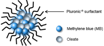
NpFlammaⓇ MB is a colloidal complex (80 ~ 100 nm) of fluorescent dye methylene blue (MB) and sodium oleate that MB-sodium oleate mixture is incorporated into self-assembled biocompatible amphiphilic Pluronic F-68 surfactant. Cationic MB dyes are electrostatically associated with anionic fatty acid to form neutral hydrophobic complex, which are encapsulated with Pluronic F-68 surfactant to produce MB nanoparticle with good colloidal stability.
MB is a clinically approved near infrared dye being used as a medication, clinical imaging dye. Due to positive charge, MB itself is difficult to penetrate the cell wall. Pluronic F-68 is FDA-approved biocompatible amphiphilic polymer surfactant being used in a variety of applications in medical field. The amphiphilic colloidal complex NpFlamma® MB selectively accumulates inside tumor cells, providing opportunities for cancer cell imaging.
| Product | Usage | Description |
|---|---|---|
cRGD Flamma® Series
|
Availability : Imaging for angiogenesis | Reacting Functionality : Fluorescent probes for the detection of angiogenesis and tumor cells |
BBBFlamma® Series
|
Availability : Imaging for brain | Reacting Functionality : Fluorescent dye penetrating blood-brain barrier |
Fucoidan Series
|
Availability : A variety of biological studies | Reacting Functionality : NIR fluorescent biopolymer |
NpFlamma® MB
|
Availability : Detection for tumor cells | Reacting Functionality : NIR fluorescent probe for cancer cell imaging |

 log
log My
My Contact